Effect of Cu and Si Wafer Substrates in Increasing Raman Signal Of Surface-Enhanced Raman Scattering-Based Au Nanoparticles
DOI:
https://doi.org/10.22452/mjs.vol44no2.8Keywords:
SERS substrates, Au nanoparticles, silicon wafer, copper waferAbstract
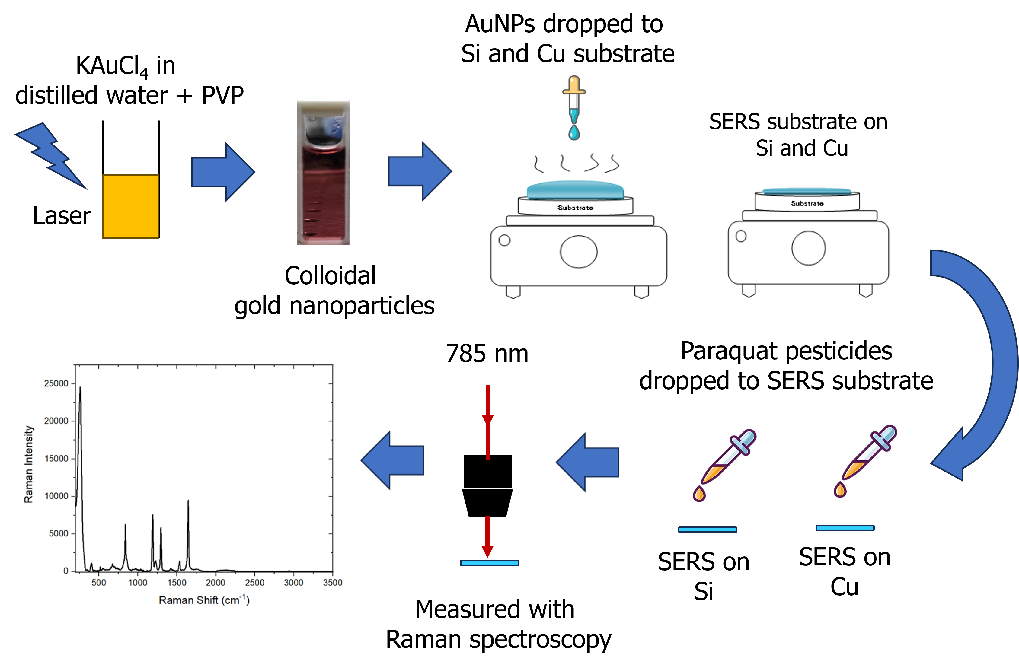
Surface-enhanced Raman spectroscopy (SERS) has attracted considerable research interest over the last four decades because of its rapid vibrational spectroscopic detection, high sensitivity, and nondestructive technique for enhancing the generally weak signal from Raman scattering. Here, SERS substrates were fabricated by drop-casting Au nanoparticles (NPs) onto two substrates (Cu and Si wafers). The AuNPs (diameter = 7.3 nm) were synthesized from an Au metal ion solution with a concentration of 4.22 × 10⁻⁴ M via photochemical reduction using a femtosecond laser. The SERS substrates were tested for their ability to enhance the Raman signal of paraquat pesticides at 10 ppm. Six vibration peaks of the paraquat pesticides at 671, 838, 1187, 1294, 1530, and 1643 cm−1 were successfully detected and enhanced. The results showed that the SERS substrate on the Si wafer increased the Raman signal more than the Cu wafer.
References
Anema J R., Li J F., Yang Z L., Ren B. and Tian Z. Q. (2011). Shell-isolated nanoparticle-enhanced Raman spectroscopy: expanding the versatility of surface-enhanced Raman scattering, Annual Review of Analytical Chemistry 4:129-150.
Chen Y T., Pan L., Horneber A., Berg M., Miao P., Xu P., Adam P M., Meixner A J., and Zhang D. (2019). Charge transfer and electromagnetic enhancement processes revealed in the SERS and TERS of a CoPc thin film, Nanophotonics 8(9), 1533-1546.
Craig A P., Franca A S., and Irudayaraj J. (2013). Surface-Enhanced Raman Spectroscopy Applied to Food Safety, Annu. Rev. Food Sci. Technol. 4:369–80.
Ferdous Z. and Nemmar A. (2020). Health Impact of Silver Nanoparticles: A Review of the Biodistribution and Toxicity Following Various Routes of Exposure, Int. J. Mol. Sci. 21(7):2375.
Gushiken N K., Paganoto G T., Temperini M L A., Teixera F S. and Salvadori M C. (2020). Substrate for Surface-Enhanced Raman Spectroscopy Formed by Gold Nanoparticles Buried in Poly (methyl methacrylate), ACS Omega 5(18):10366-10373.
Hidayah, A.N., Herbani, Y., Steven, E., Subhan, A., Triyono, D., Isnaeni, Suliyanti, M. M., and Shiddiq, M. (2022). Tuning the electrical properties of colloidal nanoalloys by varying their composition, Colloids and Surfaces A: Physicochemical and Engineering Aspects 641:128496.
Hidayah, A N., Triyono, D., Herbani, Y., and Saleh, R. (2022). Liquid Surface-Enhanced Raman Spectroscopy (SERS) Sensor-Based Au-Ag Colloidal Nanoparticles for Easy and Rapid Detection of Deltemthrin Pesticide in Brewed Tea, Crystals 12:24.
Huan Q., Liu Y., Chang S., Chen H., and Chen J. (2016). Surface Plasmonic Sensors: Sensing Mechanism and Recent Applications, Sensors 21(16):5262.
Ilyas H., Zeeshan T., Sattar N. A., Ramay S. M., Mahmood A., Abbas H. G., Saleem M. (2021). First principle and experimental investigations of monodispersed Au plasmonic nanoparticles on TiO2, Chemical Physics Letters 783:139080
Israelsen N D., Hanson C. and Vargis E. (2015). Nanoparticles Properties and Synthesis Effects on Surface-Enhanced Raman Scattering Enhancement Factor: An Introduction, The Scientific World Journal 2015: 124582.
Kahraman M., Mullen E R., Korkmaz A. and Wachsmann-Hogiu S. (2017). Fundamentals and Applications of SERS-based bioanalytical sensing, Nanophotonics 6(5)
Khalil I., Chou C M., Tsai K L., Hsu S., Yehye W A., and Hsiao V K S. (2019). Gold Nanofilm-Coated Porous Silicon as Surface-Enhanced Raman Scattering Substrate, Appl. Sci. 9: 4806.
Kneipp J., Kneipp H., Wittig B., and Kneipp K. (2010). Novel optical nanosensors for probing and imaging live cells, Nanomedicine 6(2):214–226.
Le Ru E. and Etchegoin P. (2009). Principles of Surface Enhanced Raman Spectroscopy. Pp. 134-135, UK, Oxford: Elsivier.
Lia M., Cushinga S K., and Wua N. (2015). Plasmon-enhanced optical sensors: a review, Analyst 140 (2):386–406.
Meader V K., John M G., Rodrigues C J., Tibbetts K M. (2017). Roles of Free Electrons and H2O2 in the Optical Breakdown-Induced Photochemical Reduction of Aqueous [AuCl4], J. Phys. Chem. A 121:6742–6754.
Minho K., Jung-Hoon L., Jwa-Min N. (2019). Plasmonic Photothermal Nanoparticles for Biomedical Applications, Adv. Sci. 6:1900471.
Pérez-Jiménez A I., Lyu D., Lu Z., Liu G., and Ren B. (2020). Surface-enhanced Raman spectroscopy: benefits, trade-offs and future developments, Chem. Sci. 11:4563-4577.
Pilot R., Signorini R., Durante C., Orian L. (2019). Manjari Bhamidipati and Laura Fabris, A Review on Surface-Enhanced Raman Scattering, Biosensors 9:57.
Pissuwan D., Camilla G., Mongkolsuk S., Cortie M. B. (2019). Single and multiple detections of foodborne pathogens by gold nanoparticle assays, WIREs Nanomed. Nanobiotechnol. 12:1584.
Roguska A., Kudelski A., Pisarek M., Opara M., and Janik-Czachor M. (2011). Surface-enhanced Raman scattering (SERS) activity of Ag, Au and Cu nanoclusters on TiO2-nanotubes/Ti substrate, Appl. Surf. Sci. 257(19):8182–8189.
Unser S., Bruzas I., He J. and Sagle L. (2015). Localized Surface Plasmon Resonance Biosensing: Current Chalengges and Aprroaches, Sensors 15(7):15684-15716.
Wang K., Sun D W., Pu H. and Wei Q. (2019). Shell thickness-dependent Au@Ag nanoparticles aggregates for high-performance SERS applications, Talanta 195:506-515.
Xu N., Jin S., and Li W. (2021). Metal nanoparticles-based nanoplatforms for colorimetric sensing: A review, Reviews in Analytical Chemistry 40: 1–11.
Yonzon C R., Haynes C L., Zhang X., Walsh J T., Van Duyne R P. (2004). A glucose biosensor based on surface-enhanced Raman scattering: improved partition layer, temporal stability, reversibility, and resistance to serum protein interference, Anal. Chem. 76(1):78-85.
Zhang D., Pu H., Huang L., and Sun D. (2021). Advances in flexible surface-enhanced Raman scattering (SERS) substrates for nondestructive food detection: Fundamentals and recent applications, Trends in Food Science & Technology 109:690-701.
Zhao Y., Gan S., Zhang G., Dai X. (2019). High sensitivity refractive index sensor based on surface plasmon resonance with topological insulator, Results in Physics 14:102477.
Downloads
Published
Issue
Section
License
Copyright (c) 2025 Malaysian Journal of Science

This work is licensed under a Creative Commons Attribution-NonCommercial 4.0 International License.
Transfer of Copyrights
- In the event of publication of the manuscript entitled [INSERT MANUSCRIPT TITLE AND REF NO.] in the Malaysian Journal of Science, I hereby transfer copyrights of the manuscript title, abstract and contents to the Malaysian Journal of Science and the Faculty of Science, University of Malaya (as the publisher) for the full legal term of copyright and any renewals thereof throughout the world in any format, and any media for communication.
Conditions of Publication
- I hereby state that this manuscript to be published is an original work, unpublished in any form prior and I have obtained the necessary permission for the reproduction (or am the owner) of any images, illustrations, tables, charts, figures, maps, photographs and other visual materials of whom the copyrights is owned by a third party.
- This manuscript contains no statements that are contradictory to the relevant local and international laws or that infringes on the rights of others.
- I agree to indemnify the Malaysian Journal of Science and the Faculty of Science, University of Malaya (as the publisher) in the event of any claims that arise in regards to the above conditions and assume full liability on the published manuscript.
Reviewer’s Responsibilities
- Reviewers must treat the manuscripts received for reviewing process as confidential. It must not be shown or discussed with others without the authorization from the editor of MJS.
- Reviewers assigned must not have conflicts of interest with respect to the original work, the authors of the article or the research funding.
- Reviewers should judge or evaluate the manuscripts objective as possible. The feedback from the reviewers should be express clearly with supporting arguments.
- If the assigned reviewer considers themselves not able to complete the review of the manuscript, they must communicate with the editor, so that the manuscript could be sent to another suitable reviewer.
Copyright: Rights of the Author(s)
- Effective 2007, it will become the policy of the Malaysian Journal of Science (published by the Faculty of Science, University of Malaya) to obtain copyrights of all manuscripts published. This is to facilitate:
- Protection against copyright infringement of the manuscript through copyright breaches or piracy.
- Timely handling of reproduction requests from authorized third parties that are addressed directly to the Faculty of Science, University of Malaya.
- As the author, you may publish the fore-mentioned manuscript, whole or any part thereof, provided acknowledgement regarding copyright notice and reference to first publication in the Malaysian Journal of Science and Faculty of Science, University of Malaya (as the publishers) are given. You may produce copies of your manuscript, whole or any part thereof, for teaching purposes or to be provided, on individual basis, to fellow researchers.
- You may include the fore-mentioned manuscript, whole or any part thereof, electronically on a secure network at your affiliated institution, provided acknowledgement regarding copyright notice and reference to first publication in the Malaysian Journal of Science and Faculty of Science, University of Malaya (as the publishers) are given.
- You may include the fore-mentioned manuscript, whole or any part thereof, on the World Wide Web, provided acknowledgement regarding copyright notice and reference to first publication in the Malaysian Journal of Science and Faculty of Science, University of Malaya (as the publishers) are given.
- In the event that your manuscript, whole or any part thereof, has been requested to be reproduced, for any purpose or in any form approved by the Malaysian Journal of Science and Faculty of Science, University of Malaya (as the publishers), you will be informed. It is requested that any changes to your contact details (especially e-mail addresses) are made known.
Copyright: Role and responsibility of the Author(s)
- In the event of the manuscript to be published in the Malaysian Journal of Science contains materials copyrighted to others prior, it is the responsibility of current author(s) to obtain written permission from the copyright owner or owners.
- This written permission should be submitted with the proof-copy of the manuscript to be published in the Malaysian Journal of Science
Licensing Policy
Malaysian Journal of Science is an open-access journal that follows the Creative Commons Attribution-Non-commercial 4.0 International License (CC BY-NC 4.0)
CC BY – NC 4.0: Under this licence, the reusers to distribute, remix, alter, and build upon the content in any media or format for non-commercial purposes only, as long as proper acknowledgement is given to the authors of the original work. Please take the time to read the whole licence agreement (https://creativecommons.org/licenses/by-nc/4.0/legalcode ).


